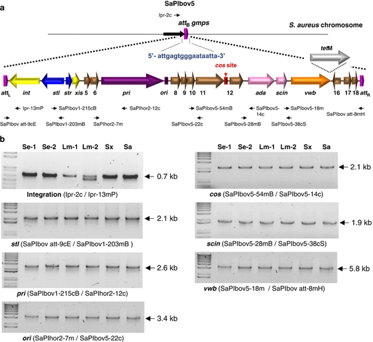
Figure 1.
(a) Map of SaPIbov5. Arrows represent the localization and orientation of ORFs greater than 50 amino acids in length. Rectangles represent the position of the ori (in purple) or cos (in red) sites. Positions of different primers described in the text are shown. (b) Amplimers generated for detection of SaPIbov5 in the different recipient strains. Supplementary Table 2 lists the sequence of the different primers used. The element was detected in S. epidermidis JP829 (Se-1), S. epidermidis JP830 (Se-2), L. monocytogenes SK1351 (Lm-1), L. monocytogenes EGDe (Lm-2), S. xylosus C2a (Sx) and S. aureus JP4226 (Sa).

