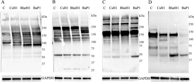
Fig 3. Western blot analysis of basement membrane components in skin homogenates.
Groups of five mice were injected by intradermal route in the ventral abdominal region with either BaP1 (PI, 75 μg), BlatH1 (PII, 1.5 μg), CsH1 (PIII, 35 μg) SVMPs or PBS (lane C). After 15 min, mice were sacrificed, their skin was removed, and an area of 12 mm diameter was dissected out. Tissues of the same group were homogenized and centrifuged, and the supernatant collected. Then, 10–20 μL of each skin homogenate sample were separated under reducing conditions on 4–15% Tris–HCl SDS-PAGE gradient gels, and transferred to nitrocellulose membranes. Immunodetection was performed with (A) anti-collagen type IV, (B) anti-collagen type VI, (C) anti-laminin, and (D) anti-nidogen 1. The anti-GAPDH antibody was used as loading control. The reaction was detected using an anti-rabbit peroxidase antibody and a chemiluminescent substrate. Images were obtained with the ChemiDoc XRS+ System (BioRad).

