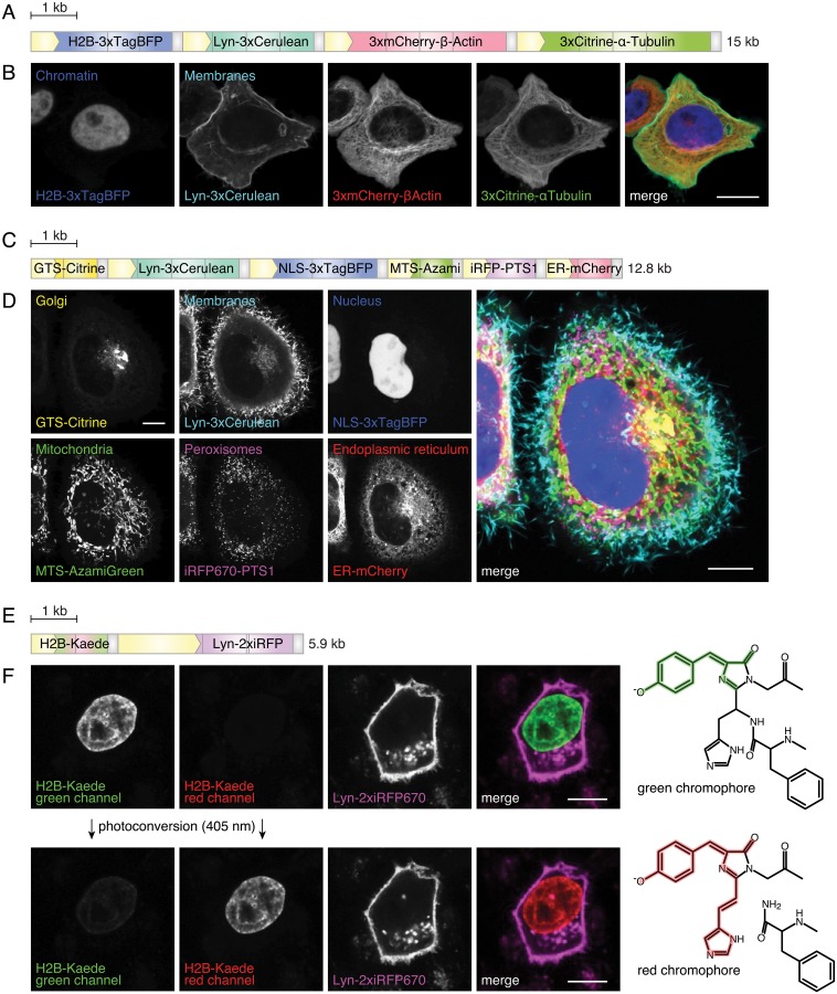
Fig 3. Tagging subcellular structures in live cells.
(A) Scheme of the construct to label nuclei, the membranes and cytoskeleton components. CMV-promoters (yellow boxes) drive the expression of trimerized fluorescent proteins fused to histone 2B (H2B), Lyn-tag, β-Actin or α-Tubulin. Gray boxes are bovine growth hormone polyAs (bGHpAs). Scale bar: 1 kb. (B) Confocal section of HeLa cells expressing the construct shown in (A). Cell organelles (nucleus and membrane) and cytoskeleton components (Actin and Tubulin) are labeled. Scale bar: 10 μm. (C) Scheme of the construct. CMV-promoters (yellow boxes) drive the expression of GTS-Citrine, Lyn-3xCerulean, NLS-3xTagBFP, MTS-AzamiGreen, iRFP670-PTS1 and ER-mCherry. Gray boxes are bGHpAs. GTS: Golgi-targeting signal, NLS: nuclear localization signal, MTS: mitochondrial targeting signal, PTS1: peroxisomal targeting signal 1, ER: N-terminal calreticulin signal peptide and C-terminal retention signal -KDEL. Scale bar: 1 kb. (D) Confocal section of HeLa cells expressing the construct shown in (C). Organelles labeled are: Golgi apparatus (Citrine), membrane (Cerulean), nucleus (TagBFP), mitochondria (AzamiGreen), peroxisomes (iRFP670) and endoplasmic reticulum (mCherry). Scale bar: 10 μm. (E) Scheme of the construct. A CMV-promoter (small yellow box) drives the expression of H2B-Kaede and a CAG-promoter (large yellow box) drives the expression of Lyn-2xiRFP670. Small gray boxes are bGHpAs, large gray boxes β-Globin polyA (β-GpA) sequences. Scale bar: 1 kb. (F) Confocal section of HeLa cells expressing the construct shown in (E) before (top row) and after (bottom row) photoconversion by a 405 nm laser. The chromophore group structures in the green- and red-emitting states are depicted on the right (adapted from [49]). Scale bar: 10 μm.

