
FIGURE 11.
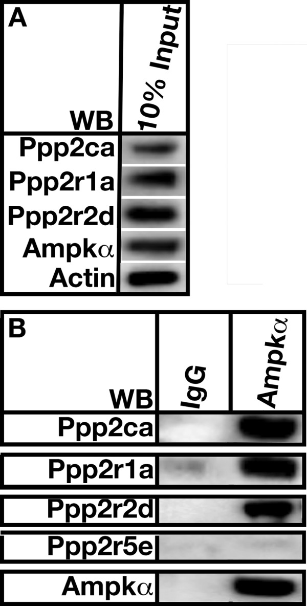
PP2APpp2r2d forms a complex with AMP kinase α in mouse aorta. A, 10% of the total protein used for co-immunoprecipitation experiments. Actin was used as a loading control. B, aortic lysate was incubated with AMP kinase antibodies (IP), and associated proteins were isolated using Protein A-Sepharose beads. Co-immunoprecipitated proteins were resolved by SDS-PAGE and detected using Western analysis (WB) using the indicated antibodies. The figure is a representation of 10 separate experiments using aortas from wild type C57BL/6 mice.