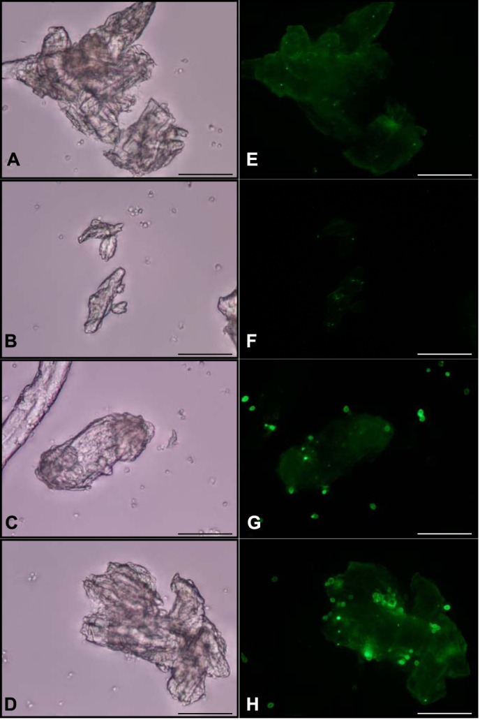
FIGURE 4.

Use of immunofluorescence microscopy to detect tāpirin proteins displayed on the cell wall of yeast. White light and epifluorescent images for S. cerevisiae EBY100 treated with anti-Calkro_0844 (A and E) or anti-Calkro_0845 (B and F) antibodies. S. cerevisiae EBY100 expressing Calkro_0844 was observed under white light (C) and epifluorescence (G) after incubation with anti-Calkro_0844 antibodies. S. cerevisiae EBY100 expressing Calkro_0845 was observed under white light (D) and epifluorescence (H) after incubation with anti-Calkro_0845 antibodies. Goat anti-rabbit conjugated with DyLight488 was used as a secondary antibody. All images were captured at ×40; scale bar in each image is 50 μm.
