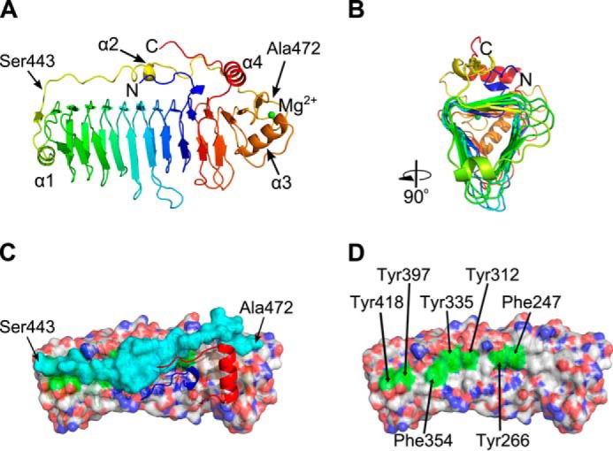
FIGURE 7.

Crystal structure of thermolysin-digested Calkro_0844_C. A, schematic representation in spectrum colors from blue on the N terminus to red on the C terminus. A single magnesium ion is depicted as a green sphere. Four α-helices are marked as well as first and last residues of the protective loop. B, cartoon representation rotated 90° to illustrate the triangular shape of the β-helix core as well as two exposed and one protected surfaces. C, view from the top onto hydrophobic surface of the β-helix core (semi-transparent surface representation, CPK colors), protective loop (semi-transparent surface, cyan), and N and C termini (cartoon, blue and red, respectively). The first and last residues of the protective loop are marked. D, view from the top onto hydrophobic surface of the β-helix core with protective loop, N and C termini removed. Exposed aromatic residues are highlighted in green and are labeled.
