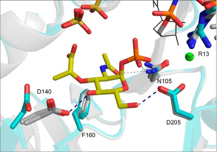
FIGURE 3.

Superposition of complex structures 1 and 2 (gray) with the native structure (cyan). The MurNAc-α1-P, UppNHp, and Mg2+ ions (representation and coloring as in Fig. 2) are shown together with a sulfate ion that is seen in all three structures. Ligands and side chains involved in ligand coordination are represented as sticks and colored by atom type (see legend to Fig. 2). Note that Asp140 is rotated by about 90° toward the C4-hydroxyl group of MurNAc-α1-P in both ligand-bound structures.
