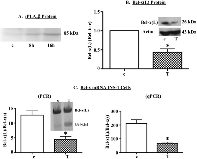
FIGURE 1.

Chemically induced ER stress correlates with reduced expression of anti-apoptotic Bcl-x(L) in β-cells. A, INS-1 cells were treated with 1 μm thapsigargin (T) or DMSO (c), and protein was extracted and used for immunoblot analysis of iPLA2β protein. A representative experiment is shown. B and C, INS-1 cells were cultured for 13 h in the presence of DMSO (c) or thapsigargin (T, 1 μm) and then RNA and protein were extracted. B, representative immunoblot analysis of Bcl-x(L) protein in c- and Tg-treated cells and quantification of three independent immunoblots. Each replicate was derived from an independent experiment that started with freshly plated cells. C, analysis of Bcl-x splice variants in a representative RT-PCR experiment (left panel inset), quantification of Bcl-x(L)/Bcl-x(S) ratio in four independent RT-PCR experiments, and quantification of Bcl-x(L)/Bcl-x(S) ratio in three independent qPCR experiments (right panel). (*, Tg group is significantly different from the c group, p < 0.05.).
