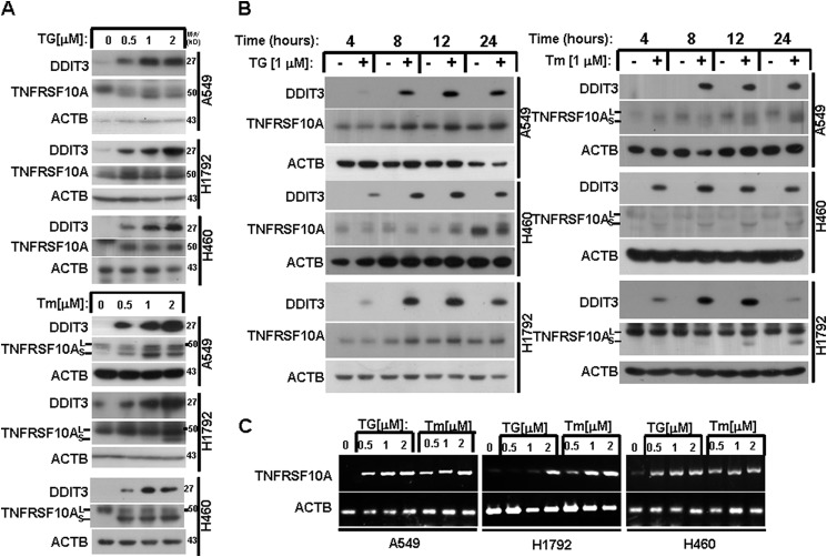
FIGURE 1.
ER stress induces TNFRSF10A expression in human NSCLC cells. A, A549, H1792, and H460 cells were treated with 0.5, 1, and 2 μm TG or Tm for 24 h. Protein expression was analyzed by Western blot using antibodies against DDIT3, TNFRSF10A, and ACTB. B, A549, H1792, and H460 cells were treated with 1 μm TG or Tm for the indicated times. Protein expression was analyzed by Western blot using antibodies against DDIT3, TNFRSF10A, and ACTB. C, A549, H1792, and H460 cells were treated with 0.5, 1, and 2 μm TG or Tm for 24 h. RT-PCR was performed to examine the mRNA levels.

