
FIGURE 5.
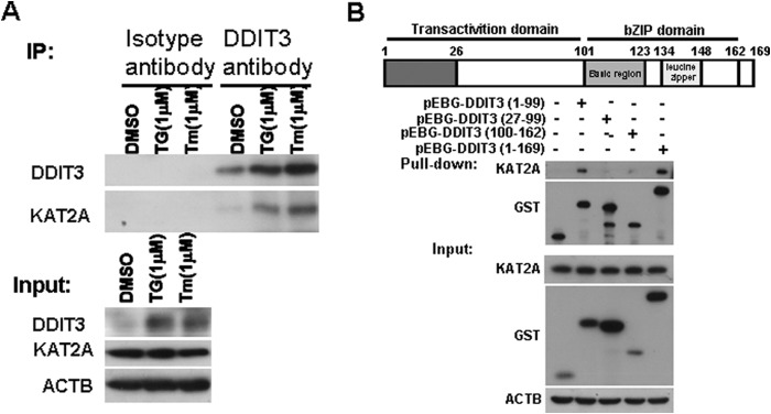
KAT2A interacts with DDIT3 by binding to its N-terminal region. A, H1792 cells were treated with DMSO, TG (1 μm), or Tm (1 μm), for 12 h, and cell lysates were immunoprecipitated (IP) using the anti-DDIT3 antibody. The isotype IgG1 antibody served as control. The proteins were detected using Western blot analysis. B, GST-tagged DDIT3 (1–99), (27–99), and (100–162) and full-length DDIT3 (1–169) were transfected into 293FT cells for 24 h, followed by glutathione-Sepharose 4B pulldown and Western blot analysis. bZIP, basic region-leucine zipper.