FIGURE 3.
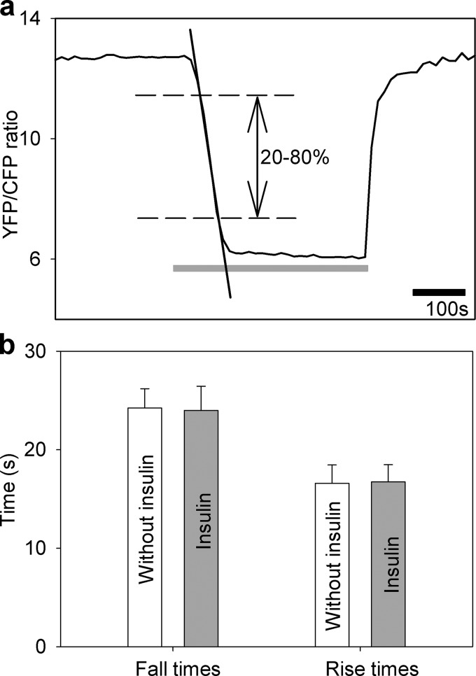
Insulin treatment does not affect the rate of glucose flux into astrocytes. a, intermittent superfusion of cells with extracellular solution containing the following d-glucose concentrations: 10 mm d-glucose for 3 min, 0 mm d-glucose for 5 min (gray line), and 10 mm d-glucose for 3 min in control untreated cells in and in cells treated with insulin (1 μm) during recordings. The time for the FRET ratio signal to rise or fall from 20% to 80% of the maximal change was determined (dashed lines), and the time interval between these two values was measured (separately for fall time20–80% and rise time20–80%). In addition, the rates of fall and rise of the FRET ratio signal were determined as indicated by linear regression line fitted to the fall of the FRET ratio signal (for details, see “Experimental Procedures”). b, fall and rise times of the FRET ratio signal in control cells without insulin (white bars, n = 10) and in insulin-treated cells (gray bars, n = 10). There was no significant difference in the fall or rise time between the two experimental settings (fall time20–80%, 24.3 ± 1.2 s (n = 10) versus 24.0 ± 1.9 s (n = 10), p = 0.91 and rise time20–80%, 16.6 ± 0.1 s (n = 10) versus 16.7 ± 1.0 s (n = 10), p = 0.92, respectively). Bars represent the means ± S.E. No differences in insulin-stimulated cells compared with control cells (fall rates were 0.16 ± 0.01 s−1 in control cells and 0.17 ± 0.02 s−1 in insulin-stimulated cells, p = 0.65; rise rates were 0.25 ± 0.01 s−1 in control cells and 0.24 ± 0.03 s−1 in insulin-stimulated cells, p = 0.69) were observed (not shown).

