FIGURE 5.

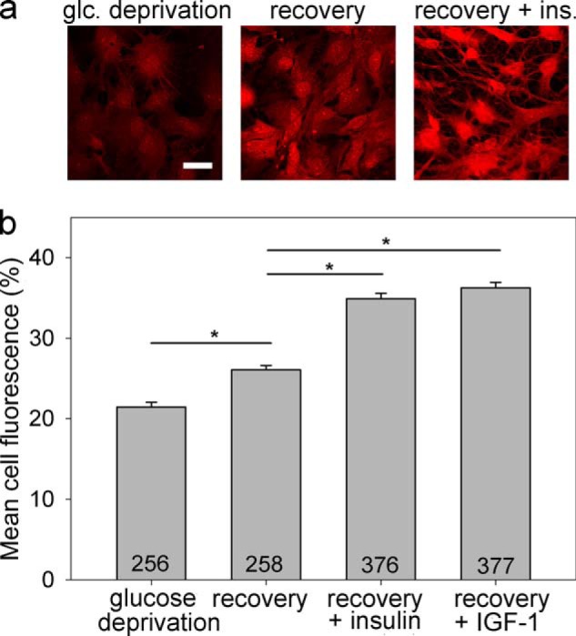
Glycogen content in astrocytes is insulin- and IGF-1-dependent. Glycogen content was determined by PAS staining. Representative images of cells positively stained with PAS (red fluorescence) after exposure to glucose-free extracellular solution for 2 h (a, left panel), glucose deprivation for 2 h, and then refeeding in 5.55 mm glucose-rich solution for 2 h (a, middle panel), and on exposure to the same experimental procedure as in the middle panel with the addition of insulin (a, right panel). The intensity of PAS staining is related to the glycogen content in the cell. Scale bar, 20 μm. b, mean fluorescence intensity of PAS staining per cell area (% of mean red fluorescence intensity per cell area over a maximal fluorescence intensity of 255 arbitrary units). Experiments were performed in cells exposed for 2 h to glucose-free extracellular solution (glucose deprivation; PAS staining: 21.4 ± 0.6% (n = 256)) after initially being exposed to glucose deprivation and then to 2 h of refeeding in the glucose-rich solution (recovery; DMEM with 5.55 mm glucose; PAS staining: 26.1 ± 0.6%; (n = 258)) and also in cells treated with either insulin (recovery + insulin; 100 nm; PAS staining: 34.9 ± 0.6% (n = 376)) or IGF-1 (recovery + IGF-1; 10 nm; PAS staining: 36.3 ± 0.7% (n = 377)) during glucose deprivation and refeeding. The numbers denote the number of cells analyzed. *, p < 0.05 (differences were tested by Kruskal-Wallis analysis of variance on ranks, and post hoc by the Dunn method); three independent experiments were performed. glc, glucose; ins., insulin.
