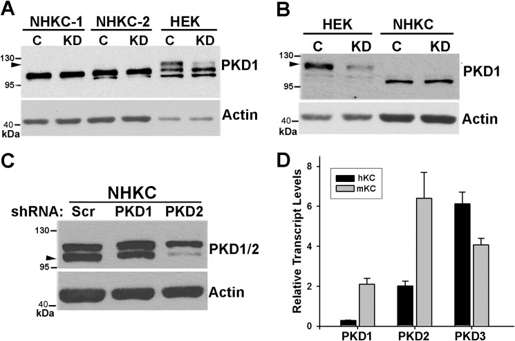
FIGURE 1.
Expression of PKD isozymes in KCs. A and B, normal human KC (NHKC) or 293HEK cells were transduced with lentiviral vectors encoding either scrambled shRNA (C) or PKD1-specific shRNA (KD). Two different strains of human KC were used in A. After drug selection, cells were lysed, and 10–30 μg of cell lysates were analyzed by Western blotting using PKD1-specific antibodies SC-639 (A) or SC-935 (B). Actin was used as a loading control. Arrowheads point to immunoreactive band representing PKD1. The position of molecular weight markers on the blot is indicated on the left. C, NHKCs were transduced with lentiviral vectors encoding shRNA against PKD1, PKD2, or a scrambled control (Scr). Protein lysates (30 μg) were analyzed by immunoblotting using an antibody cross-reacting with both PKD1 and PKD2 (CS-2052). Arrowhead points to PKD2. Actin was used as a loading control. D, quantitative RT-PCR analysis of total RNA isolated from NHKC (black bars) and primary cultures of mouse epidermis (mKC; gray bars) using primer sets specific for human and mouse PKD isoforms. Relative transcript levels after normalized to GAPDH levels are shown as mean ± S.E. of triplicates.

