FIGURE 4.
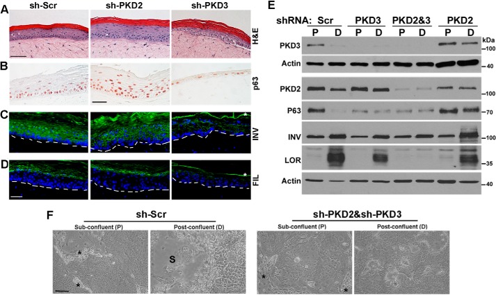
PKD3 is required for normal proliferation and differentiation in regenerating epidermis. A–D, organotypic epidermal tissue regenerated from NHKCs expressing shRNA against PKD2, PKD3, and a Scr control. Tissue sections were analyzed by histology (A) or immunostaining with antibodies that recognize pan-p63 (B; brown nuclei) or differentiation markers including involucrin (INV) and filaggrin (FIL), both in green. Blue nuclear staining is DAPI (C and D). Dashed lines in C-D indicate the position of basement membrane, and * indicates nonspecific staining of cornified layer. Scale bar, 50 μm. E, Western blot analysis of proliferating (P) and differentiating (D) cultures of NHKC stably expressing Scr-shRNA or those targeting PKD2 or PKD3, or both isoforms (PKD2&3) with antibodies that recognize PKD3, PKD2, p63, and differentiation markers involucrin (INV) and loricrin (LOR). Actin is used as a loading control. A representative of three independent experiments is shown. F, NHKCs were co-transduced with two lentiviruses encoding sh-Scr or shRNA targeted to PKD2 and PKD3 (sh-PKD2/sh-PKD3), selected in medium containing both G418 and puromycin, and grown to confluence. Phase contrast images of cultures at sub-confluence (P) and 3 days post-confluence (D) are shown. * indicates irradiated 3T3 fibroblasts in sub-confluent cultures, and S marks the area where post-confluent cultures are stratified. Bar, 100 μm.

