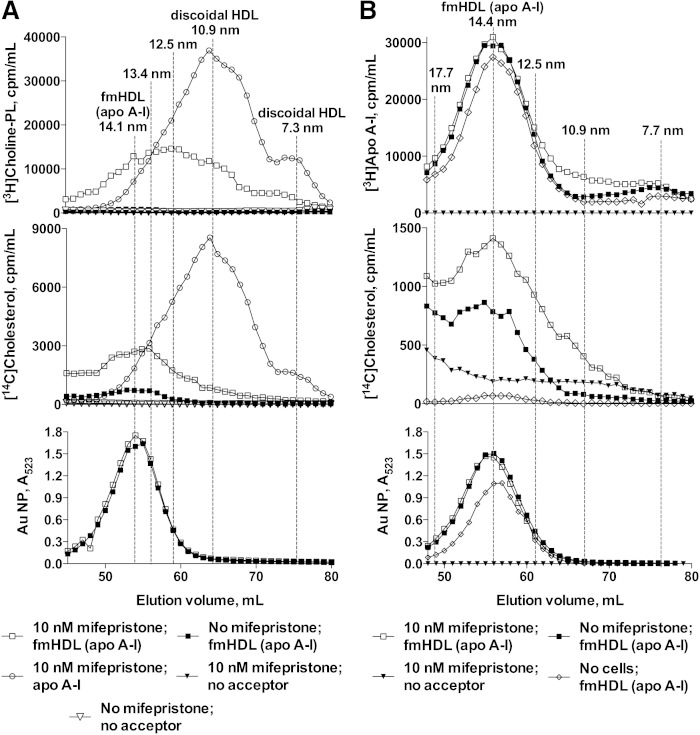
Fig. 6.
Gel filtration chromatography of fmHDL (apo A-I) exposed to BHK-ABCA1 cells with and without ABCA1 expression. A: BHK-ABCA1 cells labeled with [14C]cholesterol and [3H]choline-phospholipid were treated with vehicle or mifepristone and incubated with fmHDL (apo A-I) or lipid-free apo A-I. Cell media were concentrated and analyzed on a gel filtration column. fmHDL (apo A-I) particles were detected by measuring A523. B: BHK-ABCA1 cells were labeled with [14C]cholesterol, treated with vehicle or mifepristone, and exposed to fmHDL prepared with [3H]apo A-I. A no-cells control was generated by incubating fmHDL ([3H]apo A-I) in cell medium in tissue culture flasks without cells. The gel filtration columns used in A and B differed in the void volume. Particle sizes are approximate.

