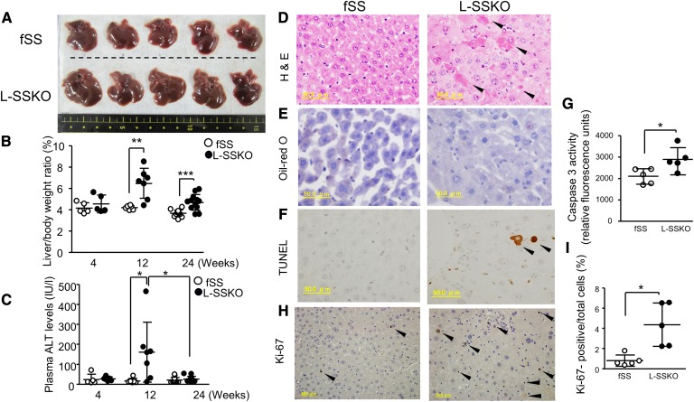
Fig. 3.
Liver-specific deletion of SS causes transient hepatic toxicity with hepatomegaly. A: Representative gross appearance of the livers from male mice at the age of 12 weeks (n = 5 in each group). Liver/body weight ratio (B) and plasma ALT levels (C) in fSS and L-SSKO male mice at the ages of 4, 12, and 24 weeks (n = 5∼13 in each group). Liver sections from male mice at the age of 12 weeks were stained with H and E (D), Oil Red O (E), and TUNEL (F). Arrowhead indicates necrotic cells in H and E staining and apoptotic cells in TUNEL staining. G: Caspase-3 activity in the livers of fSS and L-SSKO male mice at the age of 12 weeks (n = 5 in each group). The liver tissue lysates were used for this assay. H: Ki-67-stained liver sections from control and L-SSKO male mice at the age of 12 weeks. Arrowhead indicates Ki-67-positive cells. I: Dot graph shows the percentages of Ki-67-positive cells relative to the total number of cells. Each value represents the mean ± SD. * P < 0.05, ** P < 0.01, and *** P < 0.001 by Student’s t-test.

