Fig. 3.

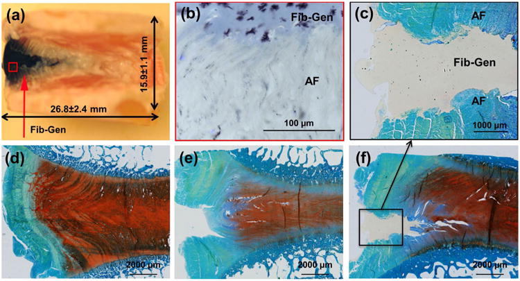
Fib-Gen repair on sagittal sections taken at Day 6. (a) Macroscopic observation of repaired IVD demonstrated that injected Fib-Gen filled the IVD defect and was easy to identify because it turned blue upon crosslinking. Some leaching of the blue cross-linked material was also present in adjacent AF tissue. (b) Microscopic imaging from cryosections demonstrated good interpenetration of Fib-Gen with the collagenous AF structure. (c-f) Methacrylate sections stained with extended FAST showed that (d) Intact IVDs exhibited strong blue staining of the organised outer AF region and strong red staining of the GAG-rich inner AF and NP regions; (e) Injured IVDs exhibited a disrupted AF with loss of GAG and IVD height; and (c,f) Fib-Gen Repaired conditions demonstrated the defect remained filled with Fib-Gen following approximately 14,000 cycles of simulated physiological loading, retained IVD height, had strong red GAG staining, and confirmed strong adhesion of Fib-Gen with the native AF tissue. No evidence for remodelling at the interface was observed (or expected) at this time point in these organ culture experiments.
