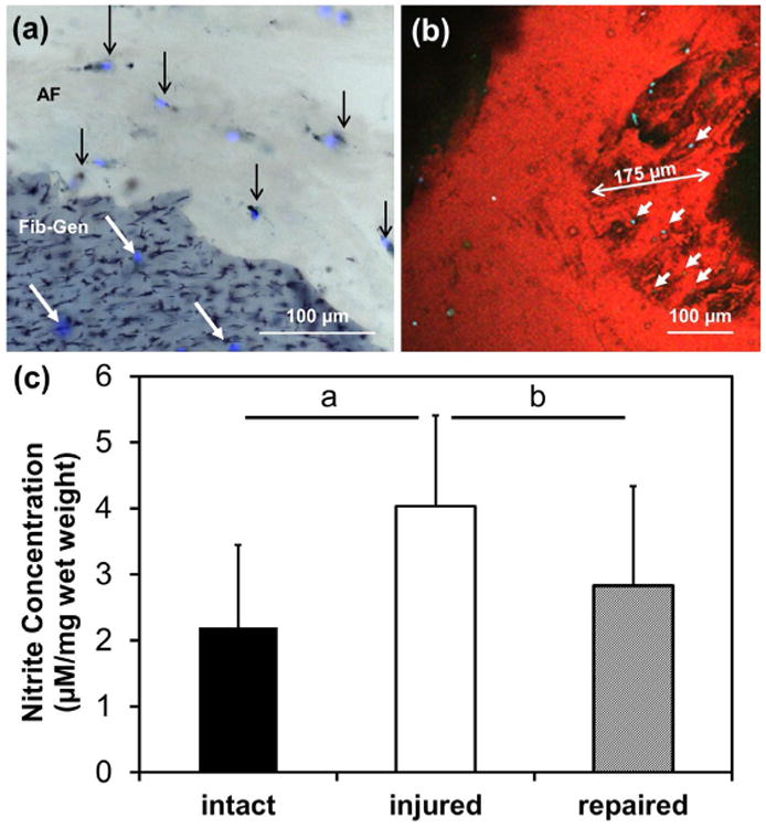
Fig. 5.

Viability imaging and NO in media. (a) Viability imaging using cryosections stained with DAPI/MTT exhibiting live cells in the AF demarcated with black arrows (live = double-stained black and blue, dead = stained for DAPI alone in blue). Migration of cells into Fib-Gen was also observed as demarcated by white arrows. (b) Confocal images further document cell migration (cyan) into the fractured interface boundary of the Fib-Gen (red colour due to autofluorescence). (c) NO levels with Fib-Gen repair were restored to intact levels. a = p < 0.01, b = p < 0.05.
