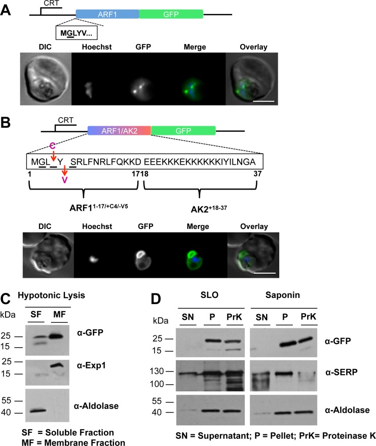
Fig 5. The subcellular location of PfARF1/GFP and GFP fused to the modified version of the N-terminus of ARF1, PfARF1-17/+4/-5AK218-37/GFP.
(A) PfARF1/GFP was expressed using the CRT promoter (construct indicated above the images) and detected by epifluorescence microscopy. PfARF1/GFP was largely located in discrete dots; Figure details as in Fig 1. (B) The chimeric PfARF1-17/+4/-5AK218-37/GFP was expressed using the CRT promoter (construct indicated above the images). Live cell imaging of the PfARF1-17/+4/-5AK218-37/GFP parasite line at the late trophozoite stage showed that the pattern of GFP expression was similar to that of PfAK2/GFP. Figure details are as in Fig 1. The PfARF1-17/+4/-5AK218-37/GFP transgenic parasite was subjected to (C) hypotonic, or (D) SLO and saponin lysis and cell fractionation as described in Fig 2. No degradation of the fusion protein was detected after Proteinase K treatment of either the SLO or the saponin pellet fractions, indicating that the protein is protected from the protease and therefore within the PPM. The labelling is as in Figs 1 and 2.

