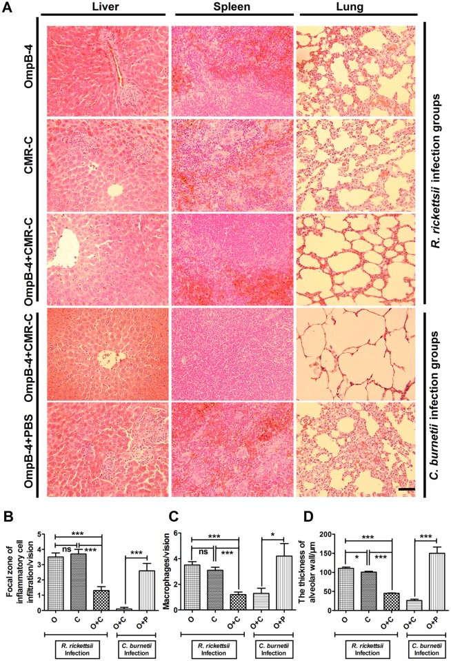
Fig 5. Pathological lesions after R. rickettsii or C. burnetii challenge.
Liver, spleen, and lung tissues were collected from mice infected with R. rickettsii or C. burnetii for pathological examination (A, original magnifications 400, bar = 200μm), respectively. The focal zone of inflammatory infiltrates in livers (B), the number of macrophage number in spleens (C), and the mean thickness of alveolar wall in lungs (D) of mice were observed. The lesions in liver, spleen, or lung were quantified (n = 10 lesion high powered fields), and the differences between the groups were compared using Student’s t-test or Wilcoxon two-sample test according to their normality and homogeneity of variance. *, P<0.05; ***, P<0.001; ns, no significance.

