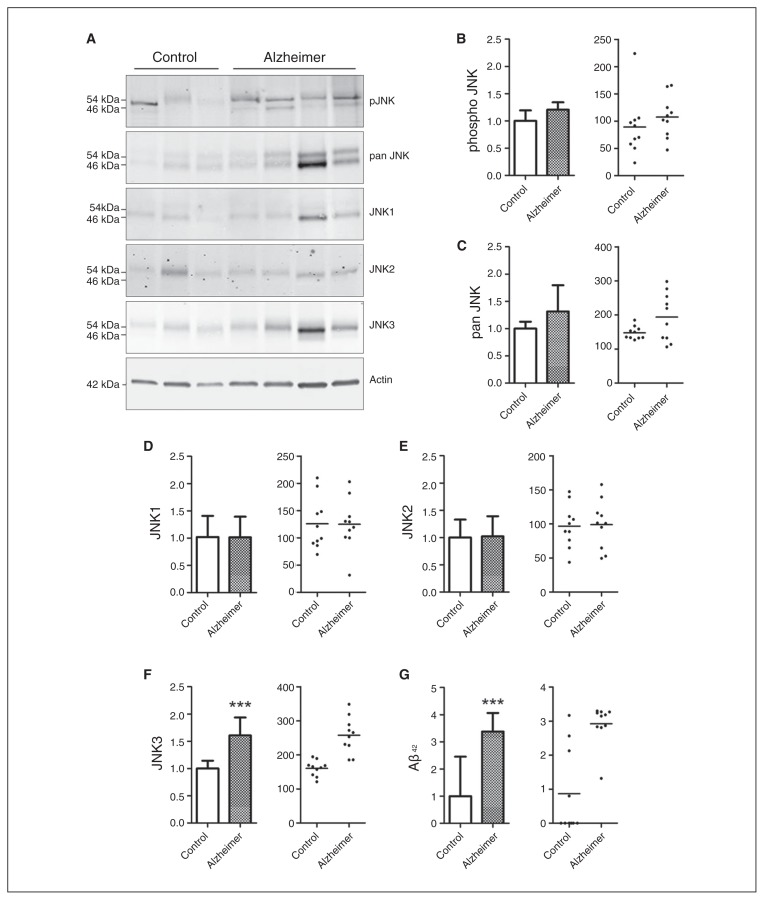
Fig. 1.
Levels of total pan JNK, pJNK, the different full form of JNK isoforms, and amyloid-β (Aβ42) in the postmortem frontal cortex of patients with Alzheimer disease (n = 10) and controls (n = 10). (A) Immunoblot analysis of pJNK, total JNK, JNK1, JNK2 and JNK3 in frontal cortex samples from patients with Alzheimer disease and controls. Corresponding histograms and scatter plots, in optical density units (ODU), of (B) pJNK, (C) total JNK, (D) JNK1, (E) JNK2 and (F) JNK3 protein levels, showing that the JNK3 isoform is significantly increased in the frontal cortex of patients with Alzheimer disease. (G) Aβ42 measured in the brain, showing an increase in the frontal cortex of patients with Alzheimer disease. ***p < 0.001. Error bars indicate standard errors of the mean.

