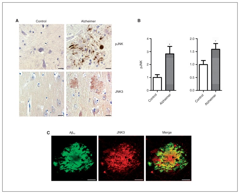
Fig. 2.
Expression and localization of pJNK and JNK3 full in Alzheimer disease and control brains. (A) Immunohistochemical studies in the frontal cortex of patients with Alzheimer disease (n = 8) and controls (n = 9) showed pJNK staining localization around senile plaques and neurofibrillary tangles (top) and JNK3 immunostaining at senile plaques (bottom). (B) The pJNK and JNK3 expression was increased in the frontal cortex of patients with Alzheimer disease compared with the same regions in controls. (C) Confocal analysis showed an association of JNK3 and amyloid-β (Aβ) labelling in senile plaques in the frontal cortex of patients with Alzheimer disease. *p < 0.05. Scale bars = 10 μm. Error bars indicate standard errors of the mean.

