
Figure 3.
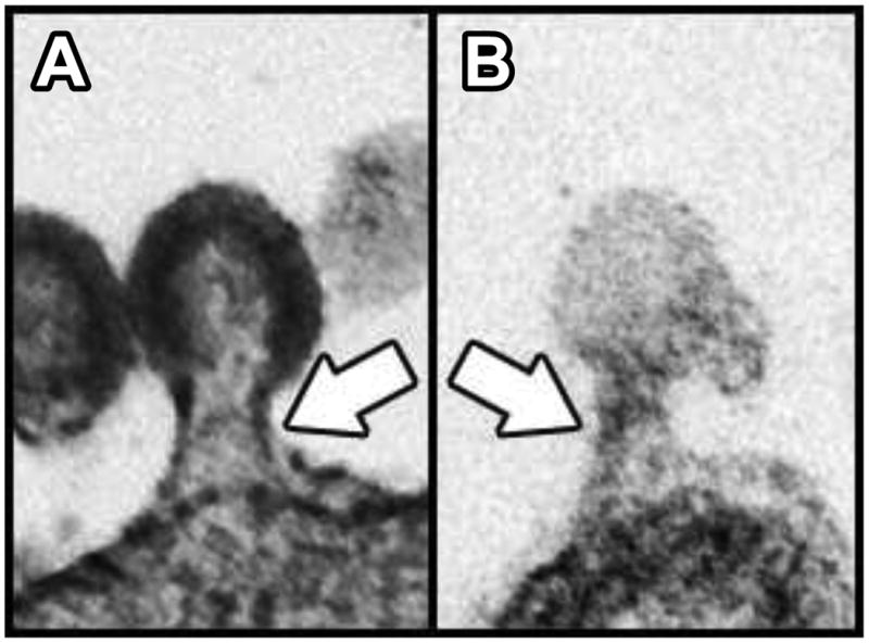
Comparison between a viral bud and a ciliary ectosomal bud. A. TEM of an ultrathin section through an HIV-1 particle arrested in the process of budding from the plasma membrane of a human cell in culture (112). B. TEM of an ultrathin section through a ciliary ectosome caught in the process of budding from the membrane of a Chlamydomonas flagellum (38). White arrows indicate the appearance of similar electron-dense structures within the two bud necks.