
Figure 5.
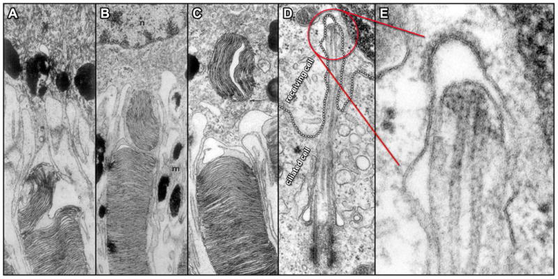
Direct interactions between cilia and neighboring cell surfaces. A – C. TEMs of ultrathin sections through three different rod outer segment tips illustrate the sequential events in the interaction with adjacent RPE cells in Rhesus monkey. A grouping of outer segment disks initially undergoes separation within the outer segment (A); followed by engulfment via RPE cell cytoplasmic extensions (B) and movement deeper into the cytoplasm of the RPE cell (C) (84). D. TEM of an ultrathin section through a fibroblast from the testes of rat shows the cell membrane of a receiving cell (emphasized by a dotted outline) forming what appears to be a coated pit in response to an intruding primary cilium extending from an adjacent ciliated cell. Panel E displays a higher magnification view of the ciliary tip region seen in D and indicated in red (125).