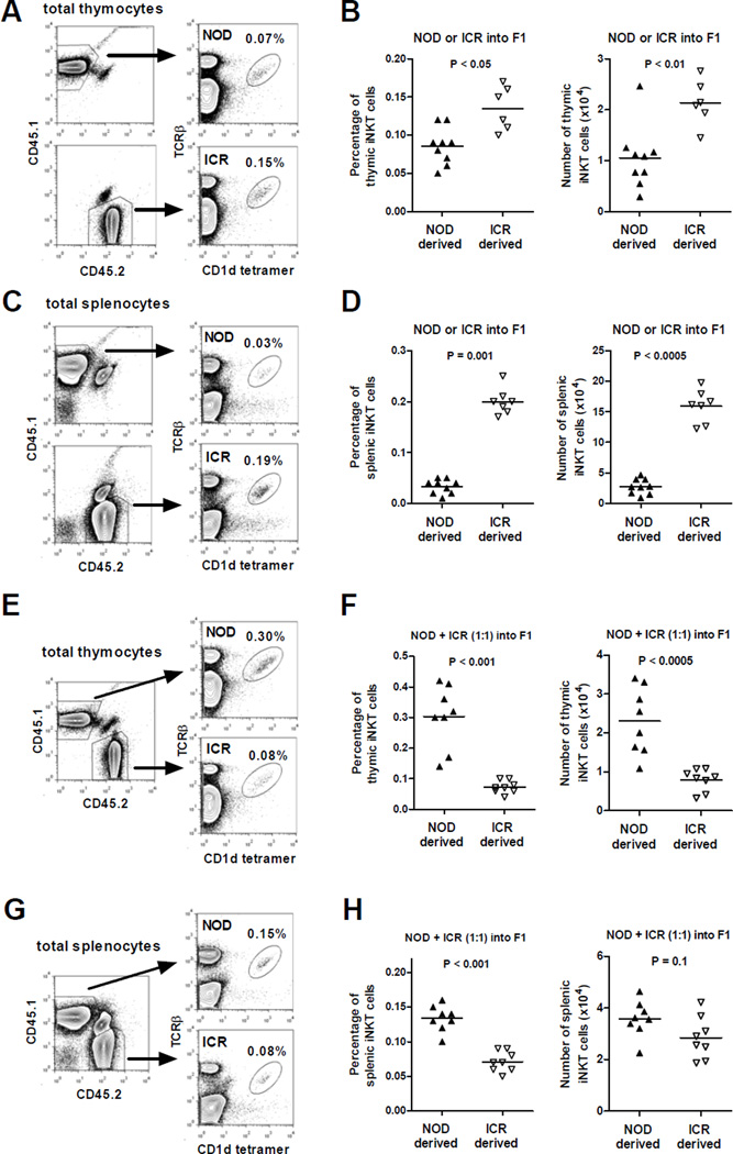
Figure 1. Hematopoietic cell intrinsic but iNKT-cell extrinsic factors contribute to impaired iNKT-cell development in NOD mice.
T-cell depleted BM cells (5×10⁶) isolated from NOD or ICR were transferred alone or co-injected at a 1:1 ratio into lethally irradiated (1100 Rads) (NOD × ICR)F1 recipients. The frequency and number of iNKT-cells were determined in the thymus and spleen at 8–10 weeks post BM reconstitution. (A and C) Representative flow cytometry profiles of the thymus (A) or spleen (C) cells isolated from an F1 recipient reconstituted with either NOD (CD45.1) or ICR (CD45.2) BM cells. (B and D) Summarized results of the frequency and number of thymic (B) and splenic (D) iNKT-cells in F1 recipients reconstituted with either NOD (CD45.1) or ICR (CD45.2) BM cells. (E and G) Representative flow cytometry profiles of the thymus (E) or spleen (G) isolated from an F1 recipient reconstituted with equal number of NOD (CD45.1) and ICR (CD45.2) BM cells. (F and H) Summarized results of the frequency and number of thymic (F) and splenic (H) iNKT-cells in F1 recipients reconstituted with equal number of NOD (CD45.1) and ICR (CD45.2) BM cells. Each symbol represents one BM recipient. The horizontal bar indicates the mean. Results are pooled from 2 independent experiments. Statistical analysis was performed using the Mann Whitney test.

