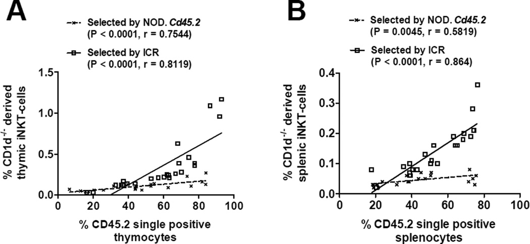
Figure 2. ICR DP thymocytes are more capable than those from NOD mice to support the development of iNKT-cells.
NOD.Cd1d−/− BM cells admixed with those from NOD.Cd45.2 or ICR at different ratios (from 4:1 to 1:4) were transferred into lethally irradiated (NOD × ICR)F1 recipients. The frequencies of NOD.Cd1d−/− derived iNKT-cells were determined in the thymus (A) and spleen (B) of the BM chimeras at 8–10 weeks post BM reconstitution. Gating strategy was similar to that described in Figure 1. The percentage of iNKT-cells is presented as the proportion of CD45.1+/CD45.2− cells. Statistically significant correlations (Pearson correlation, P < 0.05) between the percentages of NOD.Cd1d−/− derived iNKT-cells and the proportions of CD45.2+ cells were found for those selected by NOD.Cd45.2 or ICR in both the thymus and spleen. The solid and dotted lines represent the best fit linear regression lines.

