Fig. 2.
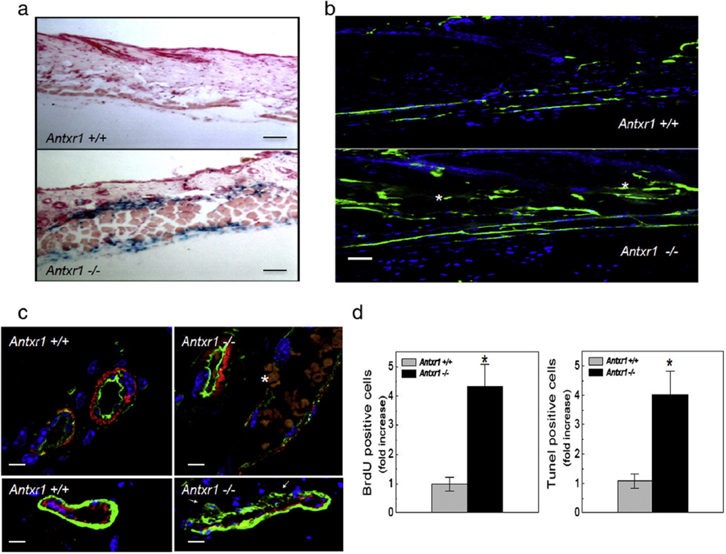
Proliferative and leaky cutaneous blood vessels in mutant mice. (a) Strong LacZ staining in cutaneous vessels (20 µm skin sections) of mutant (bottom) compared with control mouse (top) at 7 weeks. Scale bars 100 µm. (b) Immunofluorescence showing extravasation (white stars) of FITC-coupled dextran in skin section of mutant mouse (bottom) compared with control (top). Scale bars 50 µm. (c) Top: Double staining with lectin (green) and antibodies against α-SMA (red); white star at erythrocytes escaping through mutant vessel wall. Bottom: Double immunostaining for endothelial CD31 (red) and perivascular calponin (green) cell markers. White arrows indicate sites of detachment of perivascular cells from vessel wall. Scale bars 25 µm. (d) Bar graphs showing BrdU- and TUNEL-labeling data (n = 10; *P < 0.05). (For interpretation of the references to color in this figure legend, the reader is referred to the web version of this article.)

