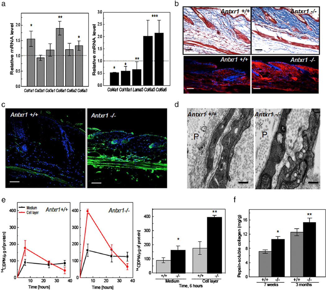
Fig. 4.
Extracellular matrix changes in mutant mice. (a) Increased transcript levels for Col1a1 and several collagen VI genes, but reduced levels for Col4a1, Col18a1 and Lama5 in skin extracts of Antxr1−/− mice (n = 6; *P < 0.05, **P < 0.005). (b) Histology (top) and immunohistochemistry (bottom) of skin sections show increased collagen deposition and increased levels of α1(I) collagen chains (red) in mutant mice. Scale bars 50 µm. (c) Immunohistochemistry shows increased deposition of collagen VI (green) in mutant skin. Scale bars 50 µm. (d) Electron microscopy indicates loss of vascular basement membranes in mutant mice. Perivascular space indicated by P. Scale bars 500 nm. (e) Left: Radiolabeling with L-[14C(U)]-Proline of collagen synthesized by fibroblasts isolated from control and mutant embryos shows increased incorporation in mutant culture medium and cell layer. Right: Differences between incorporation into collagenous protein at 6 h of incubation (n = 6; *P < 0.05, **P < 0.005). (f) Increased amounts of pepsin-resistant collagen in skin extracts of mutant mice at 7 weeks and 3 months (n = 4; *P < 0.05, **P < 0.005). (For interpretation of the references to color in this figure legend, the reader is referred to the web version of this article.)

