Fig. 5.
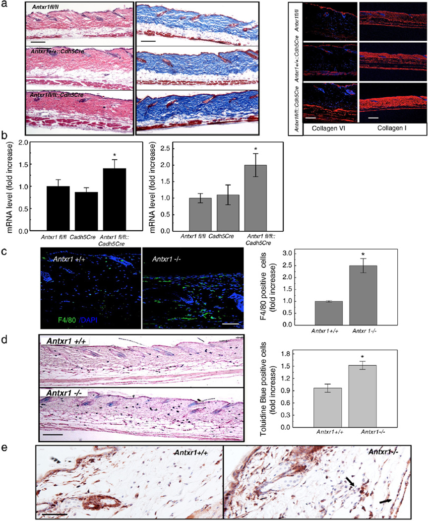
Inflammatory cells in mutant skin. (a) Histology of skin sections from control (Antxr1fl/fl and Antxr1+/+::Cdh5Cre) and homozygous (Antxr1fl/fl::Cdh5Cre) conditional knock-out mice (left and middle; scale bar 200 µm); right: immunohistochemical staining for collagens VI (scale bar 100 µm and I (scale bar 200 µm). (b) Increased levels of transcripts for Col6a1 (left, black bars) and Col1a1 (right, gray bars) in Antx1fl/fl::Cdh5Cre mutant skin (n=4; * P < 0.05). (c) Increased numbers F4/80-positive cells (green) in mutant skin. Scale bar 50 µm. (n=9; * P < 0.05). (d) Increased numbers of mast cells in mutant skin. Scale bar 200 µm. (n=10; * P < 0.05). (e) Immunohistochemical staining of skin sections for FSP1 demonstrates higher expression in cutaneous vascular regions of Antxr−/− mice. Arrows indicate FSP1-positive cells associated with vessels. Scale bars 50 µm.

