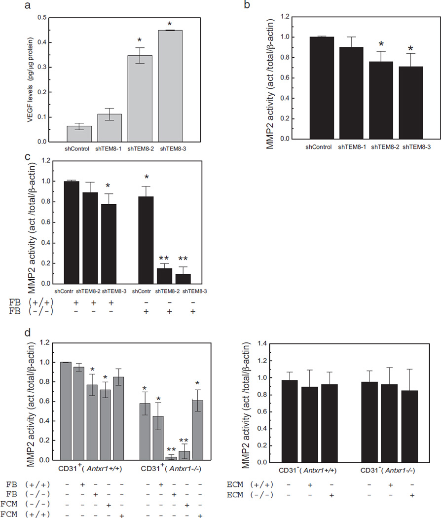
Fig. 7.
Dysregulation of MMP2 activity in TEM8 mutant cells. (a) ELISA showing VEGF levels in lysates of stable endothelial cell lines with different degrees of Antxr1 knockdown (shTEM8-1/26%; shTEM8-2/60%; shTEM8-3/90%). (n = 3; *P < 0.05). (b) MMP2 activity in lysates of stable endothelial lines with different degrees of Antxr1 knockdown. (n = 3; *P < 0.05). (c) MMP2 activity is not affected when endothelial Antxr1 knock-down cells are co-cultured with WT fibroblasts, but is dramatically reduced when the co-cultures contain fibroblasts isolated from Antxr1−/− mice. (n ≥ 3; *P < 0.05, **P < 0.005) (d) Left: MMP2 activity in lysates of primary endothelial (CD31-positive) cells from Antxr1+/+ and Antxr1−/− mice co-cultured with Antxr1+/+ or Antxr1−/− fibroblasts (FB) or exposed to conditioned medium (FCM) from such fibroblasts. Right: MMP2 activity in lysates of primary fibroblasts (CD31-negative) from Antxr1+/+ and Antxr1−/− mice cultured with conditioned medium (ECM) from Antxr1+/+ or Antxr1−/− primary endothelial cells. (n = 3; *P < 0.05, **P < 0.005).

