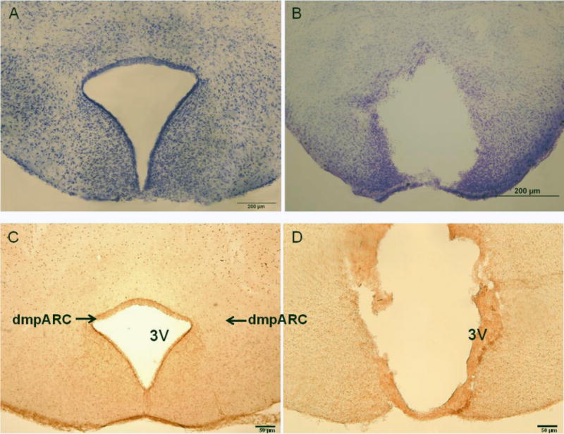
Figure 1. dmpARC Lesion Verification.

(Left) Representative microphotograph of the intact dorsomedial posterior arcuate nucleus (dmpARC) seen in the upper lateral extending corners of the ARC shown here by histamine-3 receptor immunoreactivity. (Right) A representative bilateral lesion of the dmpARC (dmpARCx) in a Siberian hamster. dmpARCsh/LD, n = 11; dmpARCx/LD, n = 5; dmpARCsh/SD, n = 13; dmpARCx/SD, n = 7. Bar: 50 μm.
