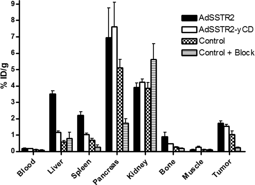
Figure 3.

Biodistribution of 64Cu-CB-TE2A-Y3-TATE in female CB.17 SCID mice implanted with MCF-7 xenografts on the rear flank. The tumors were allowed to grow with 17β-estradiol supplementation, followed by intratumoral injection of AdSSTR2 or AdSSTR2-yCD. Control mice received an intratumoral injection of saline. Two days later, 64Cu-CB-TE2A-Y3-TATE was injected intravenously into each mouse. The mice were sacrificed 4 h post-injection, and the tissues were removed to determine radioactive content. Another control group received the intratumoral saline injection followed by co-injection of 64Cu-CB-TE2A-Y3-TATE and 200 µg of Y3-TATE as a blocking agent. The data represent the mean ± SEM of the % injected dose per gram of tissue with n = 3 – 4.
