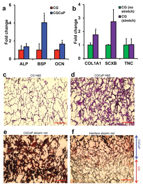
Figure 4.
MSC gene expression and histology after 6-week culture period. a) Expression of osteogenic genes alkaline phosphatase (ALP), bone sialoprotein (BSP), and osteocalcin (OCN) was elevated in the mineralized osseous compartment. b) Expression of tenogenic markers type I collagen (COL1A1), scleraxis (SCXB), and tenascin-C (TNC) was elevated in the tendinous compartment in response to tensile stimulation. Hema-toxylin and eosin (H&E) staining of the c) tendinous and d) osseous compartments revealed MSC infiltration throughout the scaffold. e) Mineral staining via alizarin red showed mineral localized primarily to the osseous compartment and f) not in the tendinous compartment. *: significantly higher expression. Scale bars: 200 μm.

