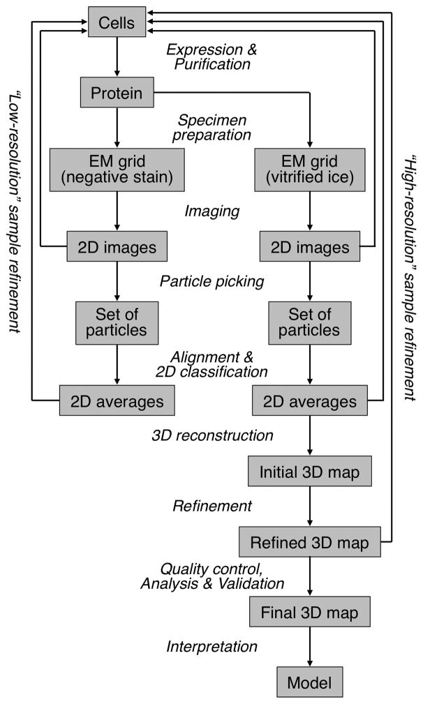
Figure 1. The Steps Involved in Structure Determination by Single-Particle Cryo-EM.
A single-particle project should start with a characterization of the specimen in negative stain (left arm of the workflow). Only once the EM images, or potentially 2D class averages, are satisfactory, i.e., the particles are mono-disperse and show little aggregation and a manageable degree of heterogeneity (“low-resolution” sample refinement), is the sample ready for analysis by cryo-EM (right arm of the workflow). The images, 2D class averages and 3D maps obtained with vitrified specimens may indicate that the sample requires further improvement to reach near-atomic resolution (“high-resolution” sample refinement).

