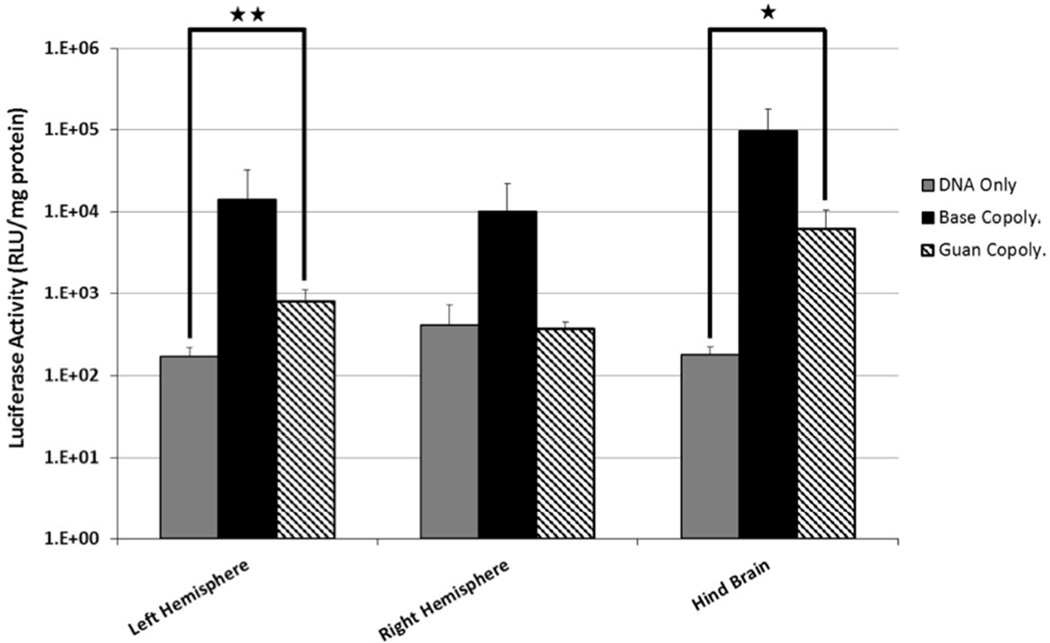
Figure 6.
Confocal micrographs of GFP+ cells 48 hrs after delivery of polyplexes containing GFP plasmid. Base Copolymer complexes injected into the lateral ventricle (yellow needle) showed Sox2-cells transfected within the ipsilateral margin (B, yellow arrows) as well a numerous cells at the contralateral ventricle margin (C). Brains injected with Guan Copolymer showed markedly fewer Sox2+ GFP+-Cells at the ipsilateral (E, cyan arrow) and contralateral margin (F, yellow). Bar, 10 µm.

