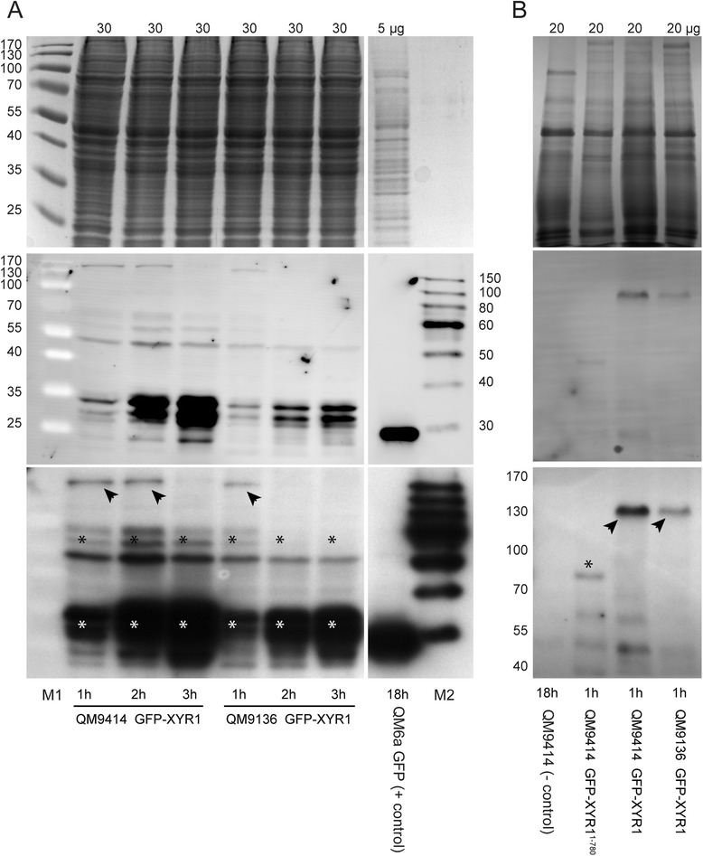
Figure 6.

Western blot analysis of GFP-XYR1 and GFP-XYR11–780 under cellulase inducing conditions. Top row: Coomassie stained loading control; middle row: chemifluorescent image of Western blot membrane; bottom row: x-ray image of Western blot membrane. (A) With time (1-3 h of sophorose induction), less of the 127 kDa full-length GFP-XYR1(arrowheads) but more of its various 30-70 kDa degradation products (asterisks) can be detected in protein extracts. This rapid turn-over of the transcription factor is conserved in QM9414 and QM9136 transformants. (B) In comparison to full-length GFP-XYR1 (arrowheads) expressed in QM9414 and QM9136 transformants, of the truncated GFP-XYR11–780 construct, only ~80 kDa or smaller degradation products could be detected (asterisk). 5-30 μg refer to the total protein load per lane; numbers on the side denote molecular weight ranges; M1 is PageRuler pre-stained molecular weight marker; M2 is SuperSignal enhanced protein ladder; 18 h QM6a GFP is the positive control, and 18 h QM9414 the negative control for αGFP(B-2) antibody specificity.
