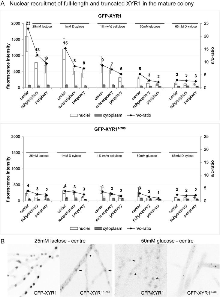
Figure 8.

Quantification of nuclear recruitment of GFP-XYR1 and GFP-XYR11–780 in the three main functional zones of the mature colony. All strains were grown for 48 hours on MA medium agar plates supplemented with the indicated carbon source. (A) Full-length GFP-XYR1 responds to different xyr1-inducing signals with nuclear recruitment, most prominently in the central area of the colony, and dependent on the inductive strength of the provided carbon source (25 mM lactose > 1 mM D-xylose > 1% (w/v) cellulose). Under non-inducing/repressing conditions (50 mM D-glucose or 65 mM D-xylose) only weak baseline nuclear recruitment can be detected. In contrast, the truncated GFP-XYR11–780 does not significantly respond to cellulase induction, and hence can only be detected at baseline levels independent of the provided carbon source. (B) Representative, inverted fluorescence images of the tested conditions, showing that GFP-XYR11–780 does respond to 25 mM lactose with weak but detectable nuclear import, comparable to the baseline presence of GFP-XYR1 under repressing conditions (barely visible nuclei are indicated with arrows). On 50 mM D-glucose GFP-XYR11–780 is visible in small spots, suggesting degradation of the non-functional XYR1 construct in the proteasome.
