
Figure 2.
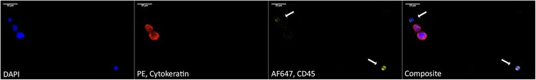
CD45 staining. Secondary staining of cell line cells spiked into healthy donor blood, from left to right: DAPI counterstain (fluorescent blue), cytokeratins 8, 18, and 19 (CK) stained with Phycoerythrin (red), CD45 stained with AlexaFluor 647 (yellow), and a composite of all channels. The two juxtaposed CTCs (CK-positive) stained negative for CD45, while the leukocytes (white arrows) simultaneously stained positive for CD45 and negative for CK, illustrating methodological selectivity.