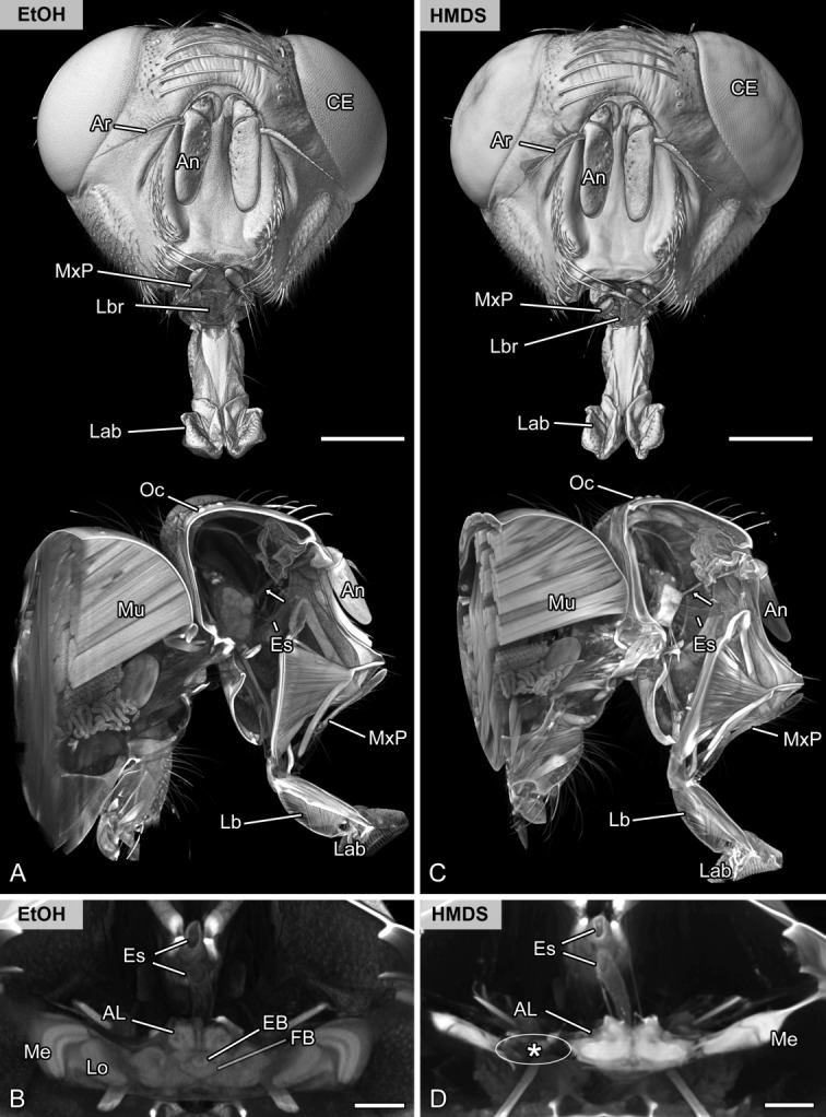
Figure 4.

Methodological evaluation of chemical drying. Volume rendering (voltex rendering) and virtual sections of the head and brain of the same specimen of the flesh fly Sarcophaga carnaria scanned in ethanol (A,B) and after chemical drying using HMDS (C,D). A,C: Frontal and oblique sagittal representation. Externally, both volume renderings show nearly equal quality. Internally, the signal to noise ratio is higher after chemical drying using HMDS (compare antennal nerve marked by arrow). However, the brain was highly affected, as indicated by enormous shrinkage artifacts. B,D: Virtual horizontal sections of the brain showing the central complex and optic neuropils. Although the signal to noise ratio is higher in the HMDS sample, internal features of the brain are not accessible. Note the high amount of shrinkage and disruption of the optic neuropils (asterisk) after HMDS treatment. Abbreviations: AL, antennal lobe; An, antenna; Ar, arista; CE, compound eye; EB, ellipsoid body; Es, esophagus; EtOH, scanned in pure ethanol; HMDS, scanned after chemically drying using hexamethyldisilazane; FB, fan-shaped body; Lab, labellum; Lb, labium; Lbr, labrum; Lo, lobula; Me, medulla; Mu, musculature of the thorax; MxP, maxillary palp; Oc, ocellus. Scale bar = 1 mm in A,C; 200 µm in B,D.
