Fig 2.

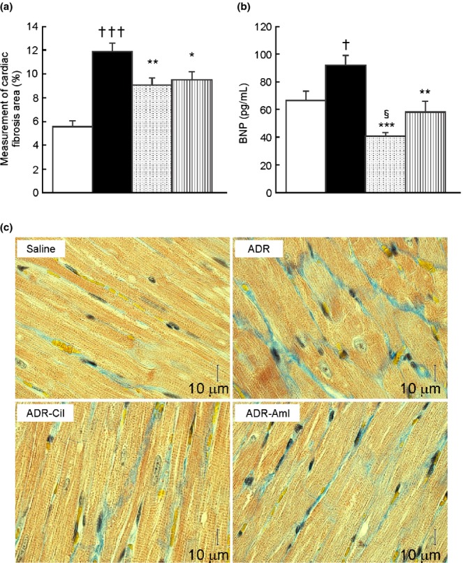
Cardiac fibrosis area (a) and plasma brain natriuretic peptide (BNP) levels (b), and micrographs of cardiac tissue with Masson's trichrome stain (c) in spontaneously-hypertensive rats (SHR) treated with adriamycin (ADR), followed by 4 weeks of vehicle (ADR group), 20 mg/kg cilnidipine (ADR-Cil), or 3 mg/kg amlodipine (ADR-Aml) administration. Saline group received saline rather than ADR, followed by vehicle for 4 weeks. Values are the mean ± standard error of the mean. †P < 0.05, †††P < 0.001 for the comparison with the saline group; *P < 0.05, **P < 0.01, ***P < 0.001 for the comparison with the ADR group; §P < 0.05 for the comparison with the ADR-Aml group.  , Saline;
  , ADR;
  , ADR-Cil;
  , ADR-Aml.
