Figure 3.
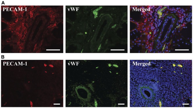
PECAM-1 is variably expressed on the inflammatory infiltrates of human minor salivary glands. Five-µm frozen Sjögren’s syndrome human minor salivary gland tissue sections were fixed as described in the Materials & Methods. Expression of PECAM-1 and von Willebrand factor was detected using immunofluorescence microscopy, as follows: mouse-anti-PECAM-1 (red); rabbit-anti-von Willebrand factor (green) and Hoechst nuclear stain (blue). Stained structures (red and green) correspond to salivary gland vasculature. The x–y cross section images were obtained using a Leica DMI6000B inverted fluorescence microscope. Presence of PECAM-1 on inflammatory infiltrates was variable, with some infiltrates expressing PECAM-1 (A), and other infiltrates not expressing it (B). PECAM-1 and von Willebrand factor were both expressed on blood vessels (A, B). Scale, 50 µm.

