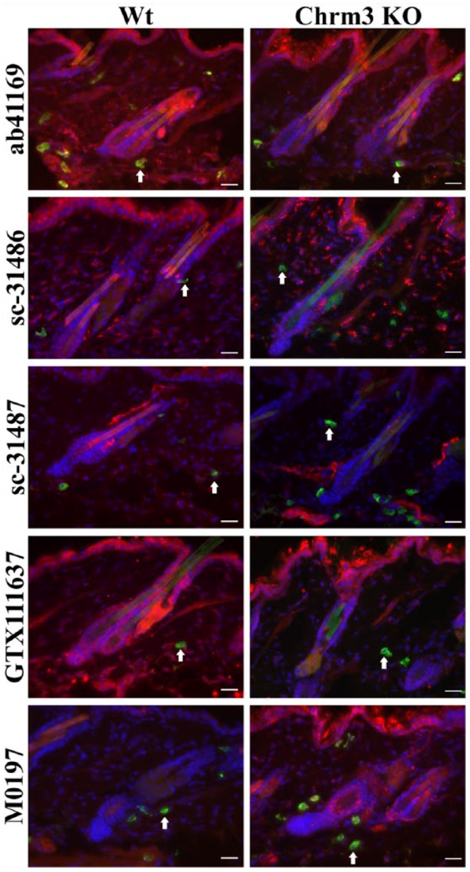
Figure 1.

Representative photomicrographs of an immunohistochemical triple staining as described in the Materials & Methods section. The stained antigen is Chrm3 (red), with staining performed using ab41169, sc-31486, sc-31487, GTX 111637 or M0194 antibodies. Mast cells (FITC-labeled avidin, green; white arrows) and cell nuclei (DAPI, blue) are counterstained. Double labelling results in a yellow signal. The left panel shows an example of the staining pattern obtained in a skin biopsy in a wild type (Wt) animal. Right panel shows the staining of skin from a Chrm3 knock-out (KO) animal. Scale, 25 μm.
