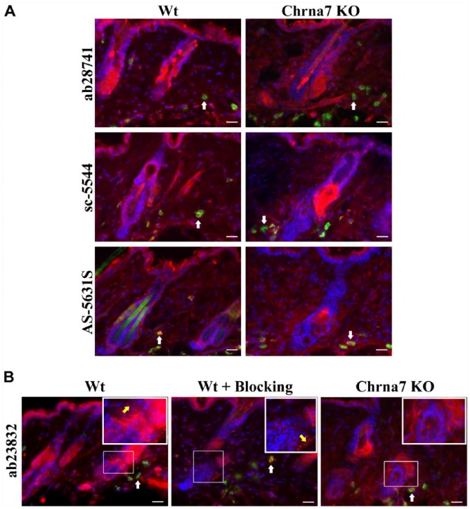
Figure 2.
Representative photomicrographs of an immunohistochemical triple staining, as described in the Materials & Methods section. (A) Stained antigen is Chrna7 (red) (ab28741, sc-5544, AS-5631S). Mast cells are FITC-labeled avidin (green, white arrows) and cell nuclei are stained with DAPI (blue). Double labelling results in a yellow signal. The left panel shows an example of the staining pattern obtained in a skin biopsy of a wild type (Wt) animal. The right panel shows the staining of skin from a Chrna7 knock-out (KO) animal. (B) Stained antigen is Chrna7 (red) (ab23832), mast cells (FITC-labeled avidin - green, pointed at by white arrows) and cell nuclei (DAPI – blue). Double labelling results in yellow signal. Regions of interest are indicated by thin white squares and shown as blow-ups in thicker white squares. Blow-ups show positive staining of nerve fibers (pointed at by yellow arrow) with ab23832 in Wt skin with or without blocking peptide (ab24285), but no positive nerve fibers in Chrna7 KO animal. Scale, 25 μm.

