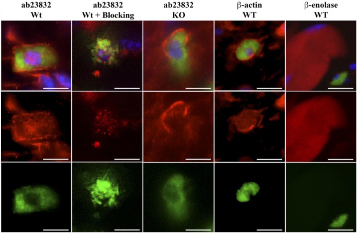
Figure 6.
Representative photomicrographs of an immunohistochemical triple staining as described in the Materials & Methods section. Stained antigens are Chrna7, β-actin or β-enolase (red) (Abcam, ab23832; Abcam; ab8227; LSBio, LS-C81180, respectively), mast cells (FITC-labeled avidin; green) and cell nuclei (DAPI; blue). First and second column panels show an example of the staining pattern obtained in the mast cells of wild type (Wt) skin without or with blocking peptide (ab24285) and the third column panels show that of skin from Chrna7 KO animals with ab23832. The fourth column panels show an example of the staining pattern of β-actin in mast cells of the Wt animal. The fifth column panels show an example of the staining pattern of β-enolase in muscle fibers of the Wt animal. Scale, 10 μm.

