
Figure 1.
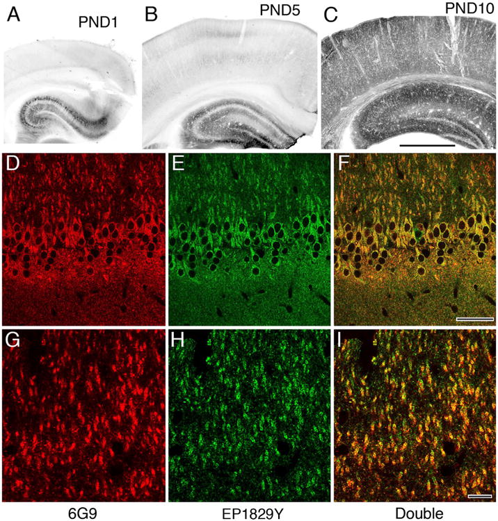
Characterization of primary antibodies used for immunohistochemistry. A,B: Early in postnatal development immunoperoxidase staining with the 6G9 antibody is weak in hippocampus (largely restricted to a few well-stained pyramidal cells), and nearly absent from overlying cerebral cortex. C: By PND 10, staining is widespread in hippocampus, and has become prominent in cortex. D–I: Immunofluorescence staining in adult CA1 for the 6G9 antibody (red channel, left) displays extensive colocalization with staining for the Origene antibody (green channel, middle), as confirmed by the yellow images on right (both channels merged). (A magenta–green version is provided as Supplementary Fig. 1 for the assistance of color-blind readers). Scale bar = 1 mm in C (applies to A–C); 50 μm in F (applies to D–F); 10 μm in I (applies to G–I). [Color figure can be viewed in the online issue, which is available at wileyonlinelibrary.com].