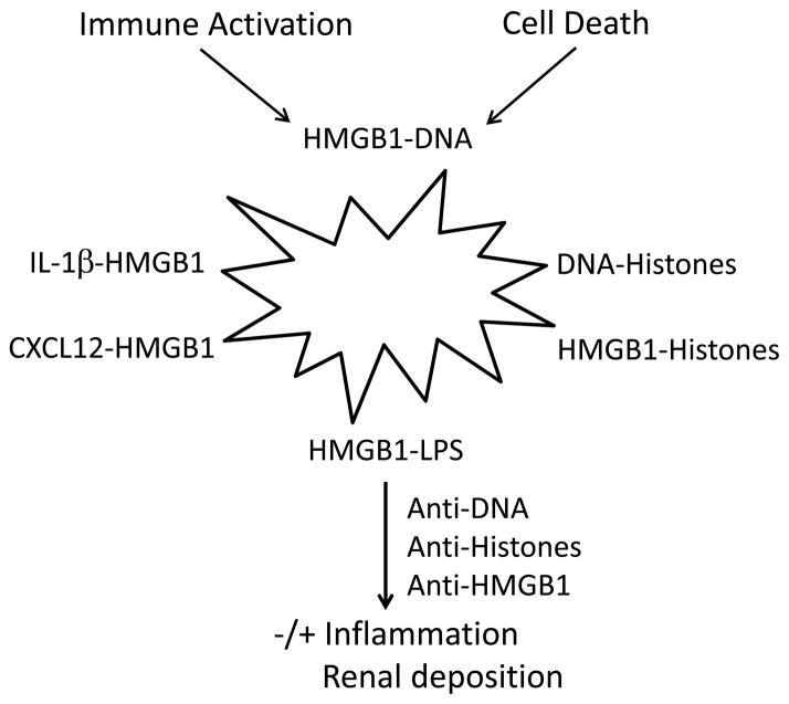
Figure 1.
The role of complexes in inflammation and autoimmunity in SLE. The figure illustrates some of the potential complexes that extracellular nuclear molecules, released during cell activation or cell deathn can form with each other as well as other immune active molecules (i.e., cytokines, chemokines or a PAMP like LPS). The cell depicted as a starburst represents a dying or activated cell, recognizing that some cells stimulated by TLR ligands undergo cell death. While all of these complexes can be formed in vitro, the existence and role in vivo for certain of these complexes in SLE, while plausible, have not yet been demonstrated. Furthermore, complexes driving events in SLE may have more components than indicated in the figure which focuses on pairs; thus, a complex of HMGB1 and LPS could also bind DNA and/or histones. These complexes in turn can interact with receptors for any of the component molecules. The figure also indicates that antinuclear antibodies can interact with complexes to stimulate or inhibit inflammation as well as deposit in the tissue.

