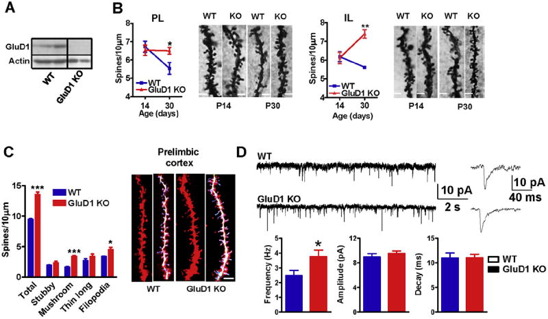
Fig. 1.

GluD1 KO mice exhibit impaired spine development and higher excitatory neurotransmission in the medial prefrontal cortex. A. Expression of GluD1 in synaptoneurosome from mPFC using western blotting. Lack of immunoreactivity demonstrates specificity of the antibody for GluD1. B. Golgi impregnation and spine analysis was performed from wildtype and GluD1 KO mice (n = 3–4 animals/age group and 15–20 neurons/animal). Impaired reduction in spine number over development was observed in prelimibic and infralimbic cortex in GluD1 KO mice with no difference in spine number between day 14 and day 30 old animals (P > 0.05, unpaired t-test). Significantly higher spine density was observed in GluD1 KO mice compared to wildtype (*P < 0.05, **P < 0.01, unpaired t-test). The bar in the image represents 2 μm. C. Diolistic labeling and spine analysis was performed from wildtype and GluD1 KO mice (4–5 animals/genotype and 15–20 neurons/animal). Significantly higher number of total spines, mushroom-like as well as filopodialike spines were found in the neurons in prelimbic cortex of mPFC of GluD1 KO mice (***P = 0.0002, ***P = 0.0001 and *P = 0.0453, unpaired t-test). The bar in the image represents 5 μm. D. Whole-cell voltage-clamp recordings from layer II–III pyramidal cells from prelimibic cortex were performed. A higher frequency of mEPSCs was observed in the pyramidal neurons in GluD1 KO mice (*P = 0.0489, unpaired t-test; N = 6 animals/group. Parameter from 2 to 3 recordings/individual animal were averaged and used for comparison.). No significant difference was observed in the amplitude or decay of mEPSCs. Recordings were performed at −70 mV in the presence of picrotoxin (100 μM) and tetrodotoxin (1 μM). Signal was filtered at 2 kHz and digitized at 10 kHz.
