


Fig. 2.
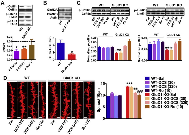
Inhibition of GluN2B-containing receptors reverses the downreguated p-cofilin-p-LIMK1 signaling and higher spine density in GluD1 KO mice. A. The levels of total and phosophorylated form of cofilin, LIMK1 and PAK1 were measured in synaptoneurosome preparation from mPFC of wildtype and GluD1 KO mice. Lower expression of p-cofilin (*P = 0.0495, unpaired t-test) and p-LIMK1 (**P = 0.001, unpaired t-test) was found in GluD1 KO mice. No difference in the expression of total proteins or p-PAK1 was observed. N = 5–8 animals/group. B. Significantly lower GluN2A/GluN2B expression ratio was observed in the synaptoneurosome from mPFC (*P = 0.0105, N = 4 animals per group). C. Mice were injected intraperitoneally with saline, DCS (30 or 320 mg/kg) or Ro-25-6981 (10 mg/kg). DCS (320 mg/kg) and Ro-25-6981 (10 mg/kg) normalized the lower p-cofilin and p- LIMK1 levels close to wildtype levels (*P < 0.05, **P < 0.01 compared to wildtype-saline, N = 4). D. Brain was collected after 30 min of systemic administration with saline, DCS or Ro-25-6981 and processed for diolistic labeling and spine analysis (4–5 animals/group and 15–20 neurons/animal). Representative images of dendrites from different treatment groups are shown. The bar in the image represents 5 μm. The total spine number was higher in GluD1 KO mice and was not significantly affected by DCS at 30 mg/kg (P > 0.05 compared to KO-saline, one-way ANOVA). Total spine density in GluD1 KO was however found to be reduced by DCS at 320 mg/kg and Ro-25-6981 (**P < 0.01 and ***P < 0.001 compared to KO-saline, one-way ANOVA).