Fig. 3.
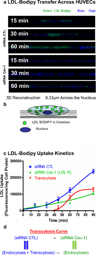
The absence of Cav-1 impairs LDL transcytosis across endothelial cells. a Visualization of cells incubated with labeled LDL from z-stacks side views (8.33 µm) across the nucleus (used as a cellular marker). b Schematic representation of z-stack side view across the nucleus as observed in a. c Quantification of LDL uptake kinetics via cellular fluorescence quantification was performed to obtain the level of transcytosis. For this experiment, cells were incubated for different amounts of time and solubilized with DMSO. Fluorescence content of each extract was measured using a fluorescence spectrophotometer. A significant reduction (57 %) of LDL internalization at 60 min was observed when cells were treated with caveolin-1 siRNA compared with the CTL siRNA-treated cells. The level of transcytosis was estimated as described in d

