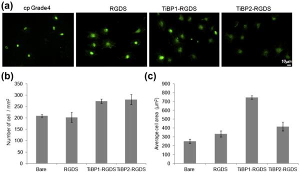
Fig. 7.
(a) FM micrographs of phalloidin stained MC3T3-E1 cells on RGDS, TiBP1–RGDS and TiBP2–RGDS-treated cp Grade 4 Ti surfaces. (b) The number of adhered MC3T3-E1 cells mm−2 in serum-free conditions on control and peptide-treated surfaces. (c) Average cell spreading on control and peptide-treated surfaces.

